Artificial Uterus Could Revolutionize Human Reproduction
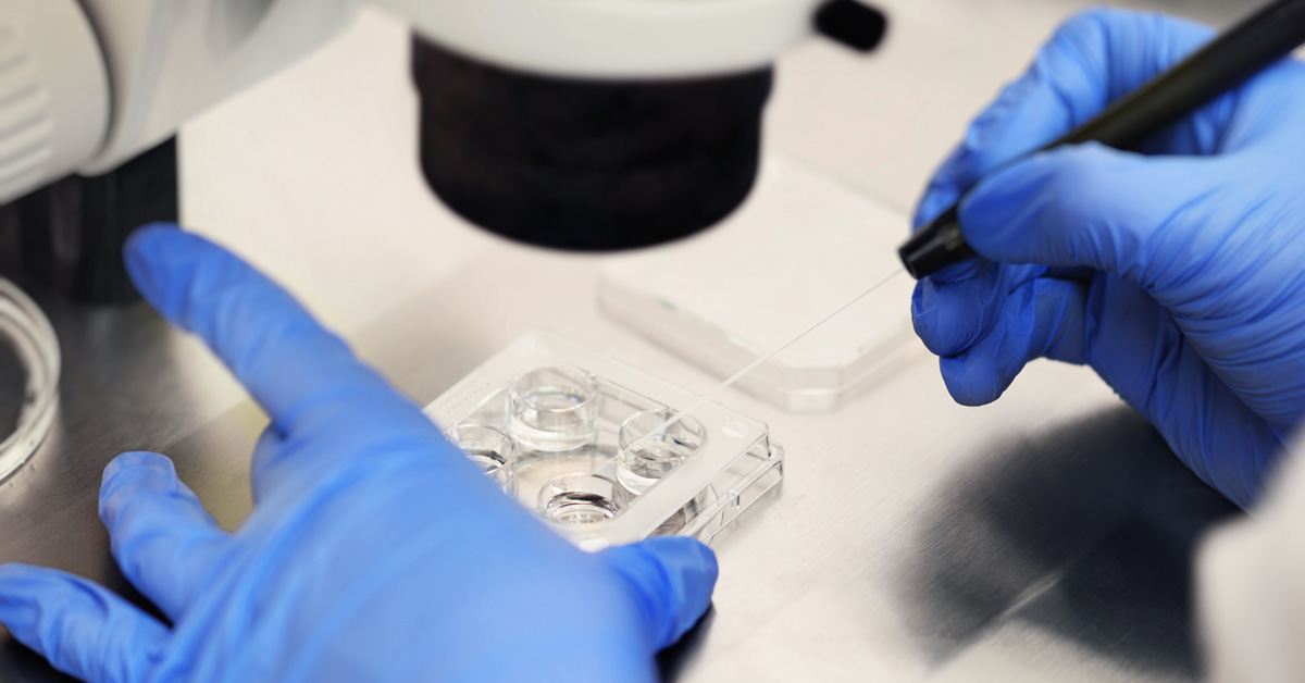
Think of a large fluid-filled sac connected to an artificial placenta. Now imagine that a premature baby is inserted inside this pouch to develop until it has sufficient health conditions to finally cuddle in its parents’ lap. Exactly that: the bag full of liquid is outside the mother. This type of experiment sounds like science fiction, but it could revolutionize human reproduction. And, if it depends on the engagement of researchers, the prototype of this premature “artificial uterus” has nothing: it should be ready within five years.
The names behind the project are designers Lisa Mandemaker and Hendrik-Jan Grievink, who, since last year, have teamed up with medical researcher Guid Oei in the search for a device that simulates the ideal conditions of the mother’s womb. It is no wonder that premature birth, that is, with the baby between 24 and 28 weeks of gestation, when, after nine months of pregnancy, it is common for the baby to be born after 39 weeks, is one of the main causes of death among babies, newborns.
According to a report from the specialized science and technology portal The Futurism, Oei, and colleagues from the Máxima Medical Center in the Netherlands obtained a donation of 2.9 million euros (around US$3.2 million, or R$12 million) to develop the prototype. “Every week, we can prolong the growth of a 24-week-old fetus in an artificial uterus; we increase the chances of survival by 18%,” explained the medical researcher. According to her, the danger of premature death probably disappears if the fetus’ development extends to 28 weeks.
Oei expects the artificial uterus to be ready within five years, although, admittedly, it is still experimental work. In other words, the consequences for the baby are not yet known. “We know nothing about the short—and long-term implications,” the researcher defined in an interview.
Not all questions have been answered, as the project is experimental. Even so, the possibility of creating an artificial prototype for the development of premature babies should represent less risk to the lives of an unimaginable number of newborns and a change in concepts about reproduction. Designer Lisa Mandemaker, for example, believes that the concepts the world has today about reproduction will have to change, especially if future technologies are challenged. In a few years, she also explained to BBC News, that an artificial uterus could be a new life choice for women.
You don’t need to worry about morning nausea, the changes in your body. There is this narrative in society that there is this ideal of natural reproduction. Natural reproduction is not the only way.
Uterus
The uterus, an important female organ related to reproduction, is pear-shaped and located in the anterior part of the pelvic cavity. The uterus is the muscular and hollow organ that is present in the woman’s body and is one of the components of the female reproductive system. This organ is fundamental for reproduction since its main function is to serve as a place for the baby’s development.
Characteristics:
The uterus is a muscular organ, with a thick wall and hollow, which is located in the anterior part of the pelvic cavity, arranged, more precisely, in the region above the bladder and in front of the rectum. A uterus, which has a shape similar to a pear, in its normal condition, is approximately 7.5 cm long and 5 cm wide (dimensions modified during pregnancy).
The uterus is a relatively mobile organ fixed by ligaments. The ligaments that ensure the fixation of the uterus are wide, round, cardinal, and uterosacral.
This organ is directly connected to the uterine tubes, which penetrate the uterus through the upper region. The two uterine tubes are also organs of the reproductive system and are characterized by being the place where fertilization generally occurs.
The uterus is where the embryo and fetus will develop, and this organ is, therefore, essential in human reproduction. After fertilization, the cilia present in the fallopian tube ensure that the embryo moves towards the uterus. The baby begins its development in this organ after implantation, which is the implantation of the embryo into the uterine wall.
When we look at the uterus, we can easily identify some parts. Are they:
- Fundus: the uppermost portion of the uterus, which has the shape of a dome. On each side of the fundus, the penetration of a uterine tube is observed.
- Body of the uterus: a region characterized by being the most dilated part of the uterus.
- Cervix, cervix, or cervix: the narrow, lower end of the uterus that communicates with the vagina. The cervix measures approximately 2 cm to 4 cm in length.
The wall of the uterus is quite developed, with three layers being observed. Are they:
- Outer layer: located more externally to the uterus and is made up of epithelial tissue.
- Myometrium: is the median and stands out for being a very thick muscular layer of smooth muscle tissue. In this layer, bundles of smooth muscle fibers are observed that are separated by connective tissue. This layer of the uterus can be further divided into three other layers, which are intertwined and, therefore, are very poorly delimited. The layer located in the middle of these layers is characterized by having a large number of blood vessels, which are essential for irrigating the organ.
- Endometrium: lines the interior of the uterine cavity, which is characterized by having a triangular shape. The endometrium can be divided into two layers: the basal layer and the functional layer. The name of these two layers already suggests their main characteristics, the first being deeper and closer to the myometrium and the second undergoing changes during menstrual cycles. It is this last part that is lost during the menstruation process. It is worth noting that after menstruation, the endometrium begins to rebuild itself through the division of new cells.
Cervix
The cervix is an important part of the uterus and, as previously mentioned, is characterized by being the lowest part of this organ. This region, unlike the rest of the organ, has a smaller amount of smooth muscle tissue, being mainly formed by connective tissue.
This part of the uterus is very important for the fertilization process, since cervical secretions are important to ensure sperm penetration into the uterus. During ovulation, secretions tend to be more fluid, which makes it easier for sperm to move.
Another important point that should be highlighted regarding the cervix is the development of cervical cancer, also called cervical cancer. This type of cancer is directly related to Human Papillomavirus (HPV) infection, and transmission is generally sexual. This cancer can be prevented by vaccination, reducing the number of sexual partners and using condoms.
Retroverted uterus
Analyzing the position of the uterus, we can say that it is anteverted or retroverted. The uterus is anteverted when it is flexed over the bladder, facing the anterior region of the body. This condition is seen in most women. In some women, however, the uterus is retroverted; that is, it is flexed backward.
A retroverted uterus can cause certain discomfort in women, who may experience pain in the lower abdomen and back, discomfort in some sexual positions, and pain during bowel movements and urination.
Curiosities
- During pregnancy, the myometrium develops due to an increase in the number and size of the cells of the smooth muscle tissue, of which it is made up.
- At the end of pregnancy, some muscle cells degenerate, and others decrease in size, ensuring the uterus returns to normal size.
- A woman’s period lasts, on average, 3 to 4 days and is nothing more than the elimination of small pieces of the endometrium and blood that are released, thanks to the rupture of vessels during the process.
- When pregnancy occurs, the menstrual cycle is interrupted.
- In some women, the presence of portions of the endometrium in an extrauterine location is observed, a condition called endometriosis. Women with endometriosis may experience pelvic pain, infertility, uterine pain, and pain during sexual intercourse.
- Cervical cancer is directly related to HPV (Human Papillomavirus) infection.
Cervical cancer, according to the National Cancer Institute, is the third most common malignant tumor in the female population (excluding non-melanoma skin cancer). This type of cancer is second only to breast and colorectal cancer.
Cervical cancer is, according to the National Cancer Institute, the fourth leading cause of death in women from cancer in Brazil.


