

Embryo Development From Fertilization to Blastocyst
Picture something invisible: an embryo growing, dividing, and changing cell by cell in the days after fertilization. Most people never see this process, yet it can influence the months that follow.
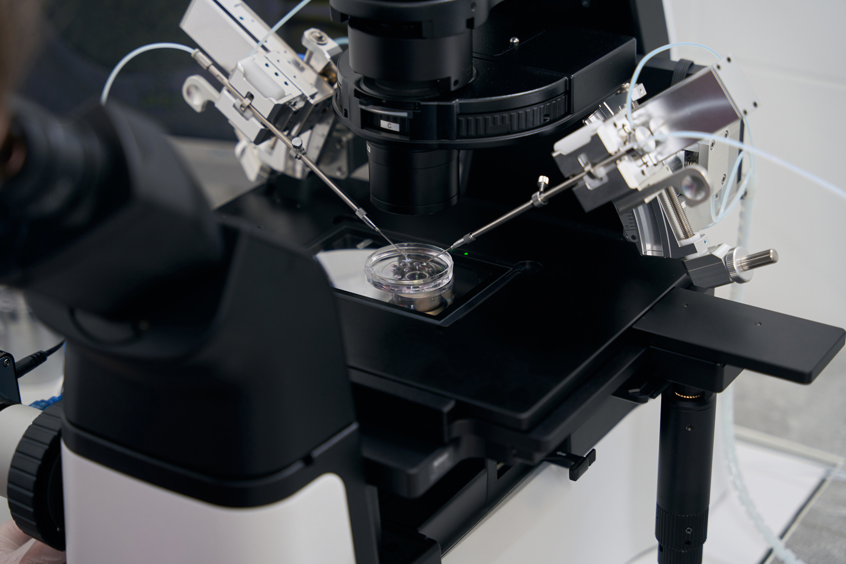
At Chedid Grieco, our laboratory in São Paulo, Brazil, follows embryos through each stage every day, using advanced reproductive technology for your in vitro fertilization treatment. Below, we walk through what happens inside the lab, step by step, so you know what our embryologists are watching for.
How Fertilization Begins
An egg is the largest cell in the human body, and it develops in the ovary long before it is needed. Once a woman reaches reproductive age, her body matures one or more eggs during each menstrual cycle, preparing them for fertilization.
The egg does not do this work alone. Sperm cells are produced continuously and are much smaller and more mobile than eggs. Their job is to travel to the egg, penetrate its outer layer, and fuse with it. When this fusion happens successfully, whether inside the body or in the lab, a new single cell called a zygote forms.
Eggs do not always have to mature inside the body first. With in vitro maturation, our team matures eggs in the lab instead. Dr. Chedid brought this technique back from her recent trip to Vietnam, and it may help patients avoid the 10 to 12 days of hormone injections usually needed before egg retrieval.
The First Five Days at a Glance
Once fertilization is complete, our embryologists check each embryo on a set schedule. Here is what they typically look for each day:
- Day 1: the zygote forms, with two pronuclei side by side, one carrying genetic material from each parent.
- Day 2: the single cell has usually divided into two, then four cells.
- Day 3: the embryo reaches around eight cells through a process called cleavage.
- Day 4: the cells compact tightly into a morula, named for its resemblance to a mulberry.
- Day 5: the embryo reaches the blastocyst stage, which contains dozens of cells.
Not every embryo follows this schedule at the same pace, which is why daily monitoring matters so much.
The Zygote and Early Cell Division
Over the next few hours, the two pronuclei merge, and the first cell division begins. Each division happens fast, and our lab checks on this progress closely, because the timing and evenness of these early splits tell us a great deal about how the embryo may continue developing.
Cleavage and Quality Assessment
By day three, an embryo has usually reached around eight cells. This is one of the most important checkpoints in the process, because not every embryo divides at the same pace or with the same symmetry.
On day three, our embryologists assess each embryo individually. These observations feed into embryo grading and selection, which helps guide decisions about transfer. Some embryos may be ready to transfer at this stage, while others benefit from a few more days of culture in the lab.
From Morula to Blastocyst
Around day four, the dividing cells compact tightly together and form a morula. This compaction is a turning point because, for the first time, the cells start forming real connections with one another rather than simply multiplying side by side.
These connections set the stage for day five, when the embryo reaches the blastocyst stage. A blastocyst contains dozens of cells and marks the final stage of laboratory development before the embryo is ready for transfer or continued monitoring. Our team supports this stage through in vitro culture of blastocysts.
Implantation in the Uterus
After a blastocyst transfer places the embryo in the uterus, the blastocyst hatches from its outer shell and attaches to the endometrium. This is why timing and lab precision matter so much for a successful blastocyst transfer.
CDC surveillance data show 85.8 percent of embryo transfers among women younger than 35 in 2021 used a single embryo. In the weeks after implantation, the embryo forms three foundational layers, the ectoderm, mesoderm, and endoderm, which give rise to every tissue and organ system in the body.
Why This Process Matters
Every embryo follows the same general roadmap, but no two cases look identical, which is why personalized monitoring matters. With fertility preservation options and lab-based techniques designed to reduce physical burden, we tailor each step to the person going through it rather than applying a one-size-fits-all protocol.
Schedule a Consultation With Chedid Grieco
Dr. Silvana Chedid leads a team of 8 doctors who have supported 8,780 babies born over 28 years of practice. Our laboratory holds FDA and NYDH licensing, including a New York tissue license held by clinics in only 8 countries outside the United States, and we perform roughly 1,200 cycles per year.
Consultations are held in Miami, while treatments are conducted at our clinic in São Paulo, Brazil, which makes fertility tourism a practical option for many families. To find out more about your own embryo’s development, contact our team to schedule a consultation.