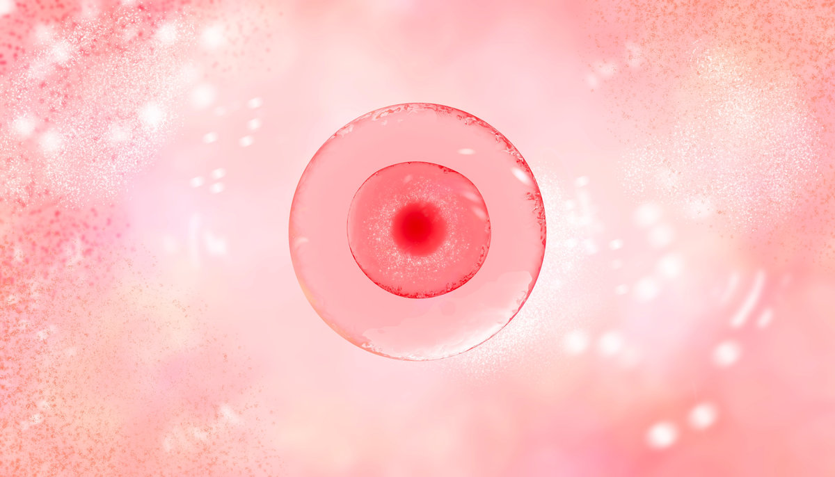
The Science Behind IVM: How Do Eggs Mature in the Laboratory?
Inside a human ovary, eggs spend months developing before they’re ready for fertilization. The natural maturation process requires precise hormonal signals and intricate cellular changes that transform an immature egg into one capable of creating new life. In Vitro Maturation replicates this biological process outside the body, allowing immature eggs to complete their development in a controlled laboratory environment rather than through weeks of hormone injections.
This approach fundamentally shifts how fertility treatment works. Instead of stimulating your ovaries with medications to mature eggs internally, embryologists retrieve immature eggs and provide them with everything needed to finish developing in culture dishes. The science behind this technique draws on decades of research into oocyte biology, growth factors, and the specific conditions that trigger the final stages of egg maturation.
What Happens During Natural Egg Maturation in the Body?
Every month, your ovaries begin developing multiple follicles during the early phase of your cycle. Each follicle contains an immature egg surrounded by support cells that provide nourishment and hormonal signals. Follicle-stimulating hormone from your pituitary gland drives this initial growth phase, causing follicles to increase in size and the eggs inside them to begin maturing.
As one dominant follicle emerges, the egg inside undergoes critical changes at the cellular level. The nucleus reorganizes its genetic material, preparing for the possibility of combining with sperm DNA. The cytoplasm accumulates proteins, energy stores, and cellular machinery needed to support early embryo development. This maturation process normally takes 10 to 14 days and culminates when a surge of luteinizing hormone triggers the final maturation steps and ovulation.
During conventional IVF, injectable hormones artificially amplify these signals to mature multiple eggs simultaneously. IVM takes a different approach by collecting eggs before this natural maturation completes, then finishing the process in the laboratory, where conditions can be precisely controlled.
How Are Immature Eggs Retrieved for Laboratory Maturation?
The retrieval process for IVM differs significantly from standard IVF egg collection. Without the 10 to 12 days of stimulation medications that conventional protocols require, your follicles remain smaller at retrieval. Ultrasound monitoring tracks natural follicle development through your regular menstrual cycle, and retrieval typically occurs when follicles reach 8 to 12 millimeters in diameter rather than the 18 to 20 millimeters targeted in stimulated cycles.
During the retrieval procedure, a thin needle passes through the vaginal wall under ultrasound guidance to aspirate follicular fluid from each follicle. The immature eggs collected through this process look visibly different from mature eggs. They have an intact germinal vesicle, the structure containing genetic material that breaks down during the final maturation steps. These eggs aren’t yet ready for fertilization, but they contain all the cellular components needed to complete development with appropriate laboratory support.
The retrieval itself takes 15 to 20 minutes and uses lighter sedation than conventional IVF retrieval because the procedure is generally less uncomfortable when follicles are smaller. Recovery happens faster as well since your body hasn’t been exposed to high-dose hormonal stimulation.
What Laboratory Conditions Allow Eggs to Mature Outside the Body?
Once immature eggs arrive in the embryology laboratory, they’re placed in carefully formulated culture media that recreate the biochemical environment inside a developing follicle. This specialized media contains specific concentrations of nutrients, growth factors, and hormones that signal the egg to continue its maturation sequence.
The culture media typically includes follicle-stimulating hormone and luteinizing hormone at precise ratios, along with growth factors like epidermal growth factor and insulin-like growth factor that support cellular development. Amino acids provide building blocks for protein synthesis, while glucose and pyruvate supply energy for the metabolically active maturation process. The media also contains serum proteins that stabilize the culture environment and protect developing eggs.
Temperature control is critical throughout the maturation period. The incubator maintains a constant 37 degrees Celsius, matching normal human body temperature. Gas concentrations are carefully regulated as well, with carbon dioxide levels adjusted to maintain optimal pH and oxygen levels calibrated to prevent oxidative stress while supporting cellular respiration. These precise conditions allow eggs to progress through their final maturation stages over 24 to 48 hours in culture.
What Cellular Changes Occur as Eggs Mature in the Laboratory?
The most visible change during laboratory maturation is the breakdown of the germinal vesicle, a large structure in the egg’s nucleus that contains genetic material. As maturation begins, the nuclear membrane dissolves, and chromosomes condense in preparation for potential fertilization. The genetic material reorganizes from a resting state into a configuration ready to combine with sperm DNA.
Simultaneously, the egg’s cytoplasm undergoes extensive reorganization. Mitochondria, the cellular structures that generate energy, redistribute throughout the cell and increase their activity. The endoplasmic reticulum, which produces proteins and lipids, reorganizes into patterns that support early embryo development. Calcium stores accumulate in preparation for the calcium wave that occurs during fertilization.
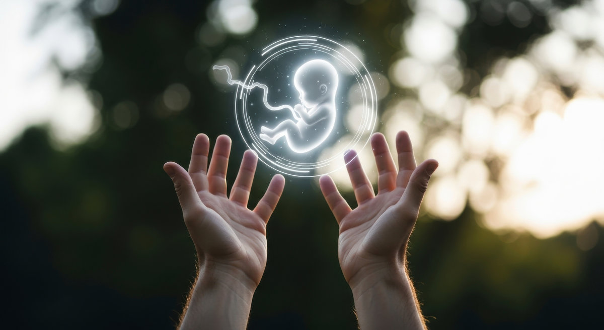
The egg also extrudes its first polar body, a small cell containing half the genetic material. This reduction from 46 chromosomes to 23 chromosomes is essential for creating an egg that can combine with sperm to form an embryo with the correct number of chromosomes. By the end of the maturation period, the egg arrests at metaphase II, the stage where it remains until fertilization triggers completion of the final cell division.
How Do Embryologists Assess Egg Maturity and Quality?
Visual assessment under high-powered microscopy provides the primary method for determining when eggs have completed maturation. Mature eggs display several characteristic features that embryologists look for during evaluation:
- Polar body presence: The first polar body appears as a small sphere between the egg and its surrounding membrane, confirming the egg has completed the first meiotic division.
- Cytoplasm appearance: Mature eggs have clear, homogeneous cytoplasm with fine granularity distributed evenly throughout the cell.
- Perivitelline space: A distinct gap appears between the egg and the zona pellucida (outer shell), indicating proper cellular organization.
- Zona pellucida clarity: The protective outer layer should appear transparent and uniform in thickness around the entire egg.
- Overall morphology: The egg maintains a round shape without fragmentation, dark areas, or other abnormalities.
Timing is crucial for achieving optimal maturity. Eggs collected at the germinal vesicle stage typically require 24 hours in culture to reach metaphase II, though some need up to 48 hours depending on their developmental stage at retrieval. Embryologists check the maturity status at specific intervals, looking for the appearance of the polar body that signals readiness for fertilization through intracytoplasmic sperm injection.
Not all immature eggs successfully complete maturation in culture. Maturation rates typically range from 60 to 80 percent, depending on factors like your age, ovarian reserve, and the specific protocol used. Eggs retrieved at more advanced stages of immaturity generally have higher maturation success rates than those collected at earlier developmental stages.
Learn More About IVM at Chedid Grieco
The science behind laboratory egg maturation represents years of research into oocyte biology and reproductive physiology. At Chedid Grieco, our FDA and NYDH-licensed facility in São Paulo, Brazil, applies this knowledge through advanced techniques refined over 30 years of reproductive medicine practice. Our embryology team has guided 8,780 families through the IVM process, carefully monitoring each egg’s development to optimize fertilization potential.
Understanding how eggs mature in the laboratory helps you make informed decisions about fertility treatment approaches. Your initial consultation happens in Miami, Florida, where our multilingual team (Spanish, English, Portuguese, and French) can explain IVM protocols in detail and determine whether this approach aligns with your medical situation and treatment preferences. Contact us today to discuss whether IVM might be right for your fertility journey. To learn more about our cross-border care model, visit our fertility tourism page.